
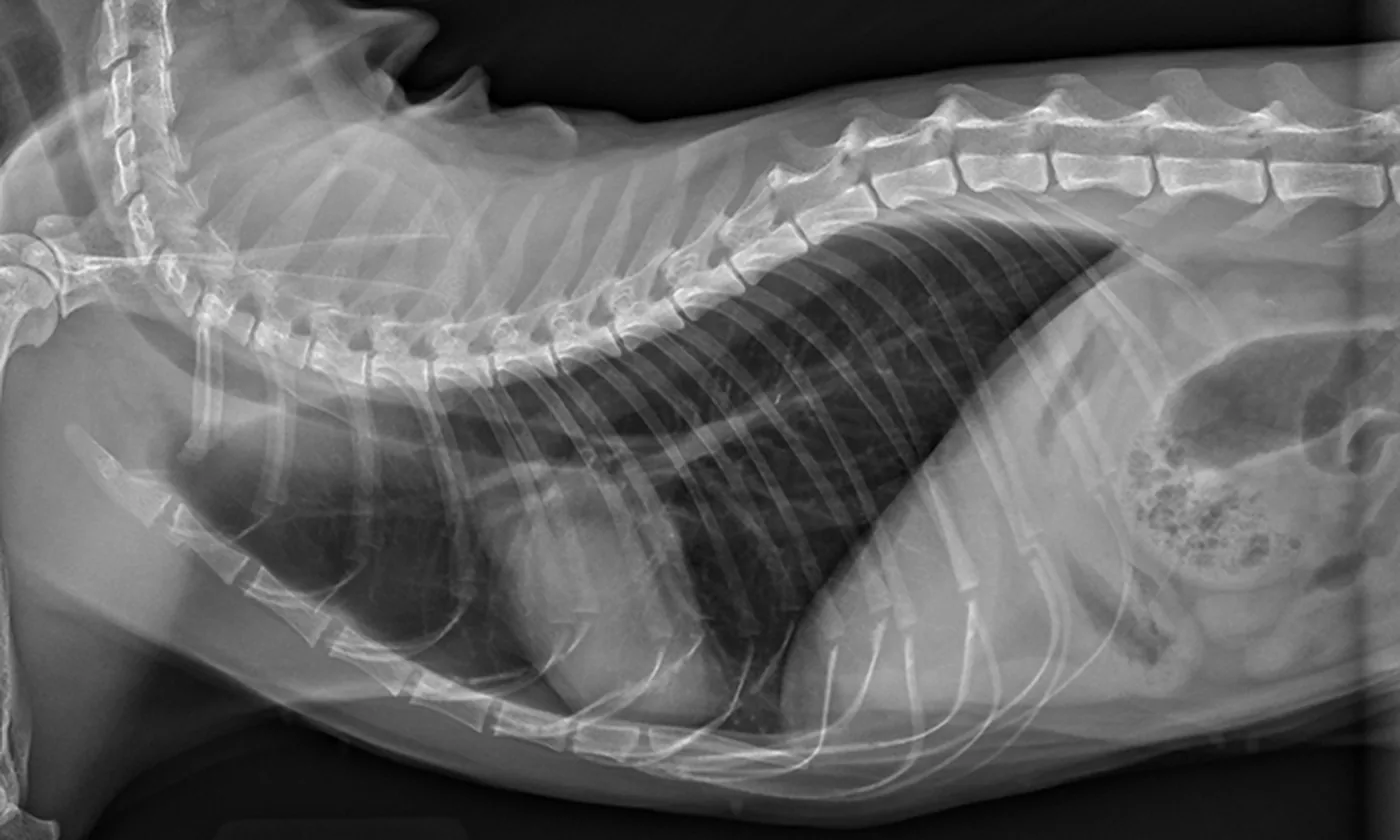
How digital x-ray may help
X-rays are particularly useful for bones and joints, the chest and patterns within the abdomen. They create a two-dimensional image, so more than one view is usually needed to understand the position and shape of structures.
X-rays cannot show every disease. Ultrasound, laboratory tests, CT, MRI or repeat imaging may be recommended when the clinical question cannot be answered with radiographs alone.
Fast digital images
Orthopaedic, chest and abdominal assessment
Referral for advanced imaging when needed
X-rays and ultrasound are complementary—not interchangeable
Radiographs are particularly useful for bones, the chest and patterns of gas or organ size. Ultrasound is better for many soft-tissue structures and movement in real time. Some patients need both because each test answers different questions.
Source: AAHA Senior Care Guidelines — Diagnostic Approach (2023)When families often book this service
- Lameness, injury or suspected fracture
- Coughing, breathing changes or heart concerns
- Vomiting, abdominal pain or suspected foreign material
- A veterinarian needs to assess the size or position of an internal structure
What your visit may include
- Several views are usually needed
- Gentle positioning is essential for useful images
- Sedation or anaesthesia may be recommended when movement, pain or stress would reduce safety or image quality
- Images are interpreted with the clinical findings and may lead to further tests
Possible tests, treatment and next steps
- The veterinarian decides which views are needed and whether calm positioning is possible while awake. Sedation or anaesthesia may improve image quality and reduce stress or pain.
- Images are interpreted alongside the examination and history. They may identify fractures, chest disease, some foreign material, bladder stones, organ enlargement or other changes.
- Complex findings may be sent for specialist radiology review or followed with advanced imaging.
Helpful things to bring or note
- Follow fasting instructions if sedation may be needed
- Tell the team if pregnancy is possible
- Bring previous images or reports when available
Going home and follow-up
- If sedation was used, follow the discharge instructions for food, supervision and activity.
- Give prescribed pain relief and restrict activity when a fracture or painful injury is suspected.
- Return promptly if breathing, pain, mobility or general condition worsens.
Digital radiography is a common first-line imaging service in general practice. CT, MRI, fluoroscopy and some interventional procedures require referral.
Major trauma, severe breathing difficulty, paralysis, a swollen painful abdomen or suspected obstruction requires prompt assessment.
Questions owners often ask
Are X-rays painful?
The imaging itself is not painful, but positioning can be uncomfortable if an area is injured. Pain relief or sedation may be used.
Why are multiple images needed?
X-rays are two-dimensional, so different angles help localise changes and reduce the chance of missing important findings.
Is ultrasound the same as X-ray?
No. They provide different information and are often complementary.
References
This information is general in nature. Your veterinarian will tailor recommendations to your pet after an examination.